Surface anatomy of the brain stem
- The brain stem is found between the diencephalon and spinal cord(bordered at foramen magnum level). It includes 3 parts:
- Midbrain中脳- also called the mesencephalon, from which it develops.
- Pons橋- develops from the metencephalon of the rhombencephalon.
- Medulla oblongata延髄- develops from Myelencephalon(髄脳) part of the rhombencephalon(菱脳,りょうのう).

Borders between the parts:
- Pons and midbrain- pons-midbrain junction (pontomesencephalic junction), made by the line between the exit of CN4(Trochlear nerve) posteriorly, to rostral edge of the basilar pons.
- Pons and Medulla:
- Ventral- Pontomedullary junction = inferior Bulbo-pontine sulcus.
- Dorsal- Striae Medullares of rhomboid fossa.
- Rhomboid fossa=floor of fourth ventricle
- Striea medullaris (carrying fibers of the arcuate nucleus) marks the inferior limit of the posterior pons
- The inside of the brain-stem is divided into 3 parts, from anterior to posterior- Basis, Tegmentum, Tectum(only in the midbrain)
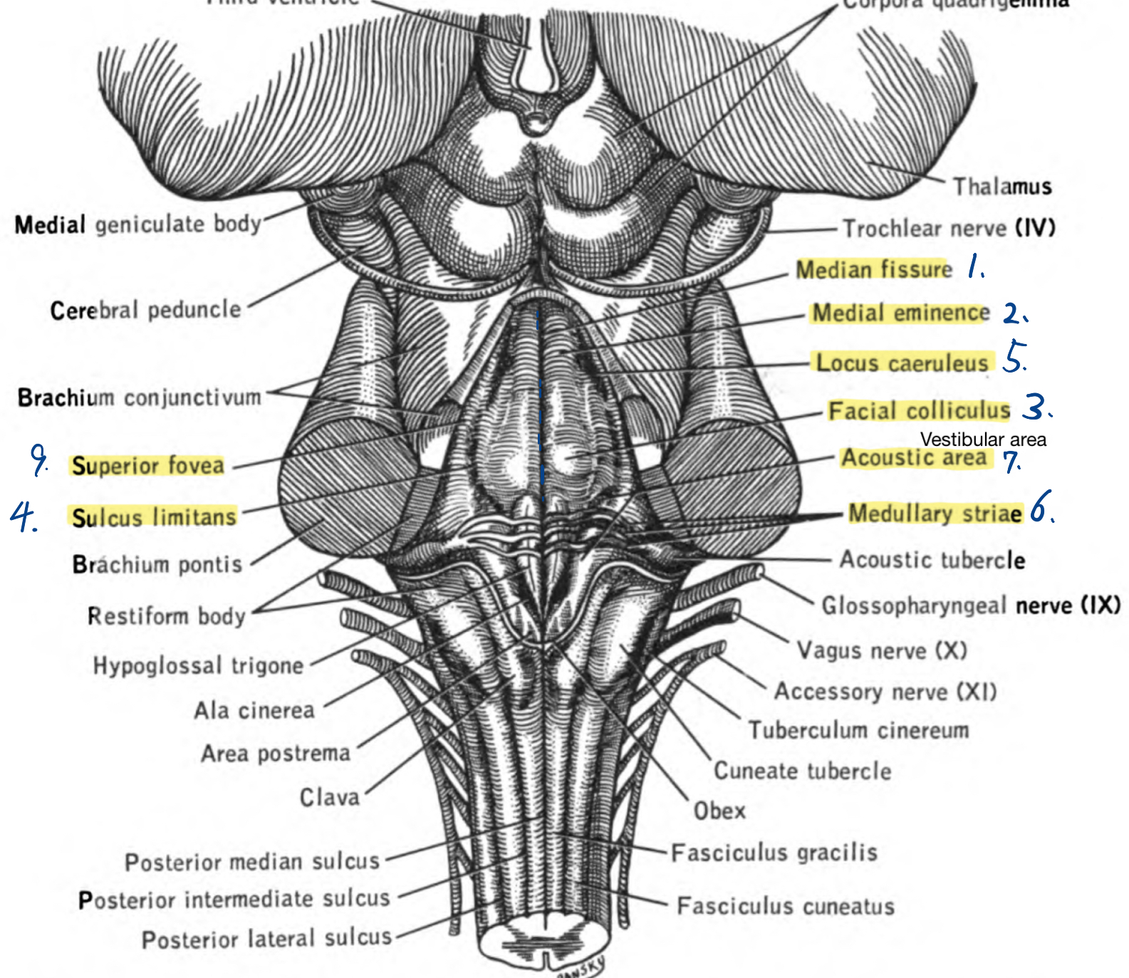
Medullary striae of the fourth ventricle
Winding around the inferior cerebellar peduncle in the lower part of the fourth ventricle, and crossing the area acustica and a medial eminence are a number of white strands, the medullary striae, which form a portion of the cochlear division of the vestibulocochlear nerve and disappear into the median sulcus.

Mesencephalon(midbrain) 1.5cm
- Location: Between the diencephalon and the pons.
- Superior border: posterior commissure and mammillary body, separate it from the diencephalon.
- Inferior border: Frenulum of the superior medullary velum
- The left colliculus of both groups is separated from its homolog on the right by a continuation of the frenulum of the superior medullary velum.
- Frenulum veli: a slightly raised white band passing from the inferior end of the medial longitudinal fissure, through the groove between the quadrigeminal bodies, and down to the superior medullary velum. On either side of this band, the trochlear never emerges and passes forward on the lateral aspect of the cerebral peduncle to reach the base of the brain.
- It has 3 parts: tectum (posterior, the colliculi, only in the midbrain), tegmentum (middle), and basis (anterior - the cerebral peduncles).
- contains the cerebral aqueduct, which interconnects the 3rd and 4th ventricles
- Supplied by superior cerebellar artery and posterior cerebral artery, branches of the basilar artery.
Ventral (anterior) surface:


- 2 cerebral peduncles - also called crus cerebri. Superiorly they enter the cerebral hemispheres, and inferiorly to the basilar pons.
- Interpeduncular fossa - between the 2 cerebral peduncles. It contains:
- oculomotor nerve (CN3) exit through this fossa, between the superior cerebellar and posterior cerebral arteries.
- posterior perforated substance - Found in this fossa, created by branches of the posterior cerebral and posterior communicating arteries penetrating and enter the brain.
- Interpeduncular cistern - Sub-arachnoid space of the interpeduncular fossa. The basilar artery passes through it. and contains the arterial circle of Willis as well as the oculomotor nerve (CN3).
- The mammillary bodies are found anterior to the interpeduncular fossa.
Dorsal(posterior) surface:

- Corpora quadrigemina - all 4 colliculi together. They form the tectum.
- Superior colliculus (SC) - Part of the visual system. The Pineal gland rests above and between them.
- Brachium of the SC - connect SC to the lateral geniculate body of thalamus.
- Inferior colliculus (IC) - part of the auditory system.
- Brachium of the IC - connect IC to the medial geniculate body of thalamus.
- Trochlear nerve (CN4) - only cranial nerve to exit from the dorsal(posterior) side of the brainstem. It exits through the border of the midbrain and pons, below the inferior colliculi.
- Quadrigeminal cistern - subarachnoid space posterior to the colliculi.
- Superior cerebellar peduncle - exit from this region.
Pons- "Bridge" in Latin. Found between cerebellar hemispheres. 2.5cm. it forms a communication pathway between the left and right hemispheres of the cerebellum.
- Location: between the midbrain and the medulla. Include the rostral part of fourth ventricle.
- extends from the superior pontine sulcus (Ponto-mesencephalic junction) to the inferior pontine sulcus (pontomedullary junction).
- It has 2 parts- tegmentum(posterior), basis (anterior).
- Blood supply- pontine branches of the basilar artery - many small branches on the anterior surface (basis) of the pons.

- Ventral surface (Anterior):
- The base of the pons - anterior part of the pons.
- Basilar sulcus - shallow groove, houses the basilar artery.
- Cranial nerves -
- Trigeminal nerve (CN5) - from the middle of the pons, little bit laterally. Marks the transition between the pons and the middle cerebellar peduncle. It has a large sensory and small motor roots.
- Abducent (CN6) - from Pontomedullary junction
- Vestibulocochlear nerve (CN8) - from the pontomedullary junction, the most laterally.
- Dorsal surface: from medial to lateral. All structures are on the floor of the rhomboid fossa (part of pontine tegmentum), covered by the superior medullary vellum:
- Median sulcus
- Medial eminence - bulges on the sides of the median sulcus.
- facial colliculus - inferior to medial eminence, contains the abducent nucleus and internal genu of the facial nerve - fibers of the motor nucleus of the facial nerve turn around the abducent nucleus.
- Sulcus Limitans - separates the alar plate from the basal plate. Between the facial colliculus and the vestibular area.
- Locus ceruleus - bluish area. contains the largest collection of norepinephrinergic neurons in the CNS. lateral to Sulcus Limitans. Respond to stress.
- Striae Medullares of the rhomboid fossa - also called Striae Acustica. The horizontal line that divides the rhomboid fossa into the superior (pontine) part and inferior (medullary) part.
- Vestibular area - partly in the pons. Overlay the vestibular nuclei.
- 3 cerebellar peduncle found on its sides. Fibers from basal pons get to the cerebellum through the middle peduncle.
- Superior fovea - lateral to facial colliculus.

Medulla oblongata - 3cm.
- Location: between the pons and the spinal cord. Include the caudal part of 4th ventricle.
- Extends from pontomedullary junction (=inferior pontine sulcus, bulbopontine sulcus) to the first cervical nerve (C1) - junction of the medulla and spinal cord, at the level of the foramen magnum.
- Supplied by branches of the vertebral artery.

- Ventral surface:
- Anterior median fissure
- Pyramid - 2 ridges on the sides of the median fissure. Contain the corticospinal tract, that passes from the pons to motor pyramidal decussation, where it crosses the midline.
- The corticospinal tract is a white matter motor pathway starting at the cerebral cortex that terminates on lower motor neurons and interneurons in the spinal cord, controlling movements of the limbs and trunk.
- Olive - also called inferior olivary eminence. Ridges on the side of each pyramid. It contains the inferior olivary nucleus. Lateral to it is the inferior cerebellar peduncle - connect medulla and cerebellum.
- 2 sulci are present, one on each side of the olive. The 4 cranial nerves of the medulla exit through three sulci
- Lateral paraolivary sulcus - also called postolivary sulcus, posterior lateral sulcus. On the lateral side of the medulla. 3 cranial nerves arise from it:
- The glossopharyngeal nerve (CN9) most superior.
- Vagus nerve (CN10)
- The accessory nerve (CN11) most inferior. Cranial part. (Actually, CN11 doesn't exit from the medulla, but from the spinal cord. These branches belong to the vagus).
- Medial paraolivary sulcus - also called preolivary sulcus and anterior lateral fissure. Found anteriorly. 1 cranial nerve arise from it: Hypoglossal nerve (CN12)
- Abducent nerve (CN6) exit through the pons - medullary sulcus, at the line of the hypoglossal nerve.
- CN7 and 8 exit posterolaterally at the pontomedullary sulcus. CN7 is more medial and it has 2 roots - motor root, and PNS root (intermediate root), which is more lateral.
- Inferior cerebellar peduncle - on the side.
- Dorsal surface: - it has 2 parts: open part - at the level of 4th ventricle, closed part - below the 4th ventricle.
- Rhomboid fossa - few structures are found at its floor, covered by the inferior vellum (superior to inferior):
- Striare Medullares of the rhomboid fossa - horizontal lines. also called striae acustica.
- Vestibular trigone - at the superior lateral corner. Overlays the vestibular nuclei.
- Inferior fovea - small depression in the limiting sulcus below the striae medullares.
- Calamus scriptorius - it consists of the hypoglossal trigone and vagal trigone. the vagal trigone is also called "ala cinerea". it is lower and more lateral relative to the hypoglossal trigone. Each covers its nucleus.
- Sulcus Limitans - separaates the alar plate from the basal plate


- Supplied by Vertebral artery system - posterior inferior cerebellar and vertebral artery and its branches (also AICA and anterior spinal artery).
- In the brain stem there are gray and white matter:
- Gray - nuclei of cranial nerve and other nuclei
- White - fibers of ascending and descending tracts from the spine, cerebellum, and cortex.


Comments
Post a Comment