Neuroanatomy study notes
Surface anatomy of the spinal cord. The spinal nerve and its roots
Gross appearance:
・shape: Cylindrical
・Borders:
-superiorly medulla of the brainstem, at the level of the foramen magnum
-inferiorly terminates at L1, in a young child it terminates at the upper border of L3. Its fibers continue downward as cauda equina.
- It occupies the upper 2/3 of the vertebral canal. At the beginning of the development, they are equally long but the vertebrae grow faster, so the vertebral canal is longer.
- Surrounded by the meninges(髄膜): dura(硬膜), arachnoid(クモ膜) & pia mater(軟膜), and CSF(脳脊髄液) in the subarachnoid space(クモ膜下腔).
- Cervical and lumbar enlargements (intumescence): bigger anterior horn of gray matter(灰白質), because it gives nerves to limbs through the brachial and lumbosacral plexuses
- Conus medullaris(脊髄円錐): inferior end of the spinal cord, becomes narrow like a cone of ice cream, at the level of L1-L2 in adults.
- Filum terminale internum(内終糸): prolongation of the pia mater inferiorly from the apex, attaches to the end of dura mater. The dura attaches the coccyx- filum terminale exernum.
- Denticulate ligament(歯状靭帯): formed by pia mater. From each level o the cord, about 20-22 small strings adhere laterally to arachnoid and dura mater. They stabilize the cord.
- Cauda equina: inferior to the end of the spinal cord, lumbar, and sacral spinal nerve continues downward. found in the Lumbar Cisterne (subarachnoid space). Cauda equina is a collection of spinal nerves and nerve roots. (Latin: Horse's tail) The cauda equina nerves control bladder and bowel, sexual function, sensation in the groin area, and motor function throughout the legs.
The inside structure of the spinal cord:
- Composed of the inner core of the gray matter, surrounded by the white matter, the segmentation is not seen on gross appearance.
Gray matter(灰白質):
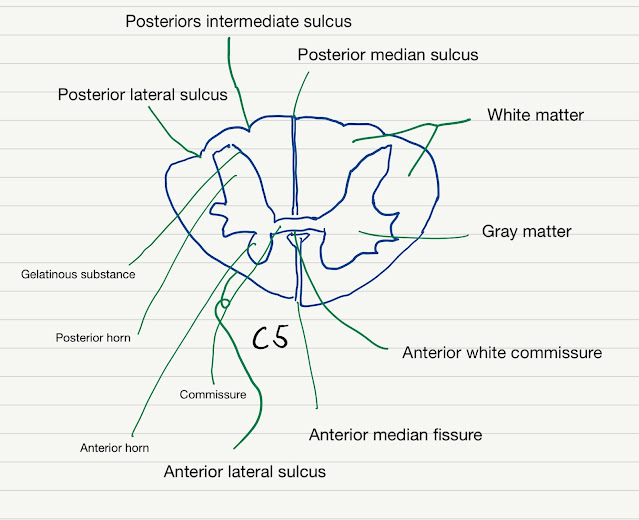
- H-shaped pillar with anterior and posterior horns (columns) united by a thin gray commissure in the middle, in which a small central canal is found. The anterior horn is motor, and the posterior horn is sensory. Each is divided into few histological laminae.
- The amount of gray matter is related to the amount of muscle innervated at that level. For that reason, there are cervical and lumbosacral enlargements, that give nerves to the upper and lower limbs.
- the central canal is found in the center of the grey matter, It is filled with CSF and continues superiorly with the 4th ventricle(第四脳室)
- Ventral horn(前角)- motor neurons(SM). It is narrow at the thoracic(胸部) level and enlarged at cervical(頸部) & lumbar(腰部) level (due to the plexuses)
- Lateral horn(側角)- only at the level of T1-L3, visceromotor(VM) neurons, that give preganglionic myelinated fibers that pass through white communicating rami to the sympathetic ganglia.
- Dosal horn(後角)- somatosensory (SS) and Viscerosensory(VS)
The gray matter is a mixture of nerve cells and their processes, neuroglia, and blood vessels. The nerve cells are multipolar.
2. White matter
- Through it, the axons ascend or descend.
- Descending pathways(下行経路): Axons that descend from the brain to the spinal cord. These are efferent fibers that affect the target tissue.
- Ascending pathways(上行路): Axons that ascend from the spinal cord to the brain. These are afferent fibers that send information from sensory cells to the brain.
- The axons of these pathways pass through one of 3 areas, called funiculus- posterior, lateral, and anterior funiculi
- Posterior funiculus=脊髄後索
- lateral funiculus=脊髄側索
- anterior funiculus=脊髄前索
The external surface of the spinal cord: On the surface of the cross-section we can see few sulci:
Posteriorly- 3 sulci. They are close, so it helps to identify the posterior part of the spinal cord:
- Posterior median sulcus- not deep
- Posterior intermediate sulcus- on the 2 sides of the posterior median sulcus, Between gracile and cuneate.
- Posterior lateral sulcus (dorsolateral sulcus)- the entrance of dorsal root (sensory fibers) from DRG. The posterior spinal artery passes medial to it.
Anteriorly-
- Anterior median fissure- deepest groove on the cord. House the anterior spinal artery.
- Ventrolateral sulcus- exit point of the ventral root.
The spinal nerve: 31 pairs of spinal nerves: 8 cervical, 12 thoracics, 5 lumbar, 5 sacral, 1 coccygeal
- Each spinal nerve has afferent and efferent fibers, coming from 2 different roots:
- Anterior root gives the efferent (motor) fibers. These fibers send information from CNS to effector cells. Their cell bodies are found in the anterior horn of spinal gray matter.
- Posterior root, gives the afferent (sensory) fibers carry information from the PNS to CNS. Their cell bodies are found in the dorsal root ganglion.
- The spinal nerve is formed by posterior and anterior roots, which are the collection of anterior/ posterior rootlets exiting from the spinal cord.
- The roots unite at the intervertebral foramen to form the spinal nerve. Then the spinal nerve divides into anterior and posterior rami that innervate different structures.
- Functionally: afferent and efferent functions have somatic (conscious) and visceral (autonomic, unconscious) components. So in total, there are 4 types of fibers- GSA, GVA, GSE, GVE (General Somatic Afferent 一般体性求心性/ General Visceral Afferent 一般内臓求心性/ General Somatic Efferent 一般体性遠心性/ General Visceral Efferent 一般内蔵遠心性)
- General Visceral Efferent 一般内蔵遠心性 get to preganglionic autonomic ganglia.
- Special fibers(SSE, SVA) involve the cranial nerves and not the spinal cord.
- Dermatone- the area of skin that is supplied by one spinal nerve.
Fibers in the posterior root
General Somatic Afferent (proprioceptive)
sensory information arising from tendons, muscles, or joint capsules
General Somatic Afferent (exteroceptive)
Pain and temperature sensation coming from the surface of the body
General Visceral afferent (interoceptive)
it conducts sensory impulses (usually pain or reflex sensation) from the internal organs, glands, and blood vessels to the central nervous system.
Cross-section of the spinal cord:
- The first thing to notice is overall shape, the cervical sections tend to be oval (wide and squashed), and the lumbar section is round.
- The ventral horn is enlarged. At segments that control a limb, the motor neurons are large and numerous, this causes enlarged ventral(anterior) horns in two places: the lower cervical sections (C5~C8) and the lumbar/sacral sections.
- The amount of white matter relative to gray matter decreases as you move down the cord. This is logical, in the white matter of the cervical cord you have all of the axons going to or from the entire body. In the sacral cord, the white matter contains only the fibers that go to inferior parts of the body. That is why the sacral cord looks like it has so much gray matter - it has lost all of the white matter.
- Cervical- wide flat cord, lots of white matter, ventral horn enlargements.
- Thoracic- have a lateral cell column. Also called an intermediate horn. These are pointed tips between the small dorsal and ventral horns. From it, the sympathetic nerves are distributed to the body. Those are found only in T1-L2/3.
- Lumbar- Round cord, ventral horn enlargements.
- Sacral- Small round cord, almost no white matter.


